Understanding The Heart
To help you understand your cardiac procedure, we have provided an overview of the normal functioning of the heart.
- The heart is a muscle, located in the left central area of your chest, behind the sternum (breastbone) and ribs. It is about the size of an adult’s closed fist and weighs less than a pound.
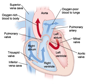
- There are four chambers in the heart, two on the right and two on the left. The upper two chambers are called atria and the lower two chambers are called ventricles.
- The main function of the heart is to pump blood to the body. More specifically it acts as two separate pumps. The right side of the heart pumps blood to the lungs, where the blood receives oxygen. The left side of the heart pumps oxygen-rich blood to the entire body through the aorta.
Normal Conduction of the Heart
Each heartbeat begins when a special group of cells located in the right atrium of the heart, known as the Sinus Node, sends an electrical signal. This signal spreads throughout the atrium to the atrioventricular (AV) Node. The AV Node connects to a special group of conducting fibers in the ventricles. As the electrical impulse travels through the heart, the heart contracts, and pumps blood to the lungs or to the body. This normally occurs 60-100 times each minute. Each contraction represents one heartbeat.
Coronary Arteries
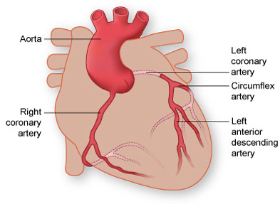
The heart, along with the rest of the body, must have oxygen to do its job. The special blood vessels that supply the heart with oxygen-rich blood are called coronary arteries.
They are located on the surface of the heart. There are two primary coronary arteries, the left coronary artery and the right coronary artery. Each of these arteries has smaller branches that also work to supply the heart with oxygen.
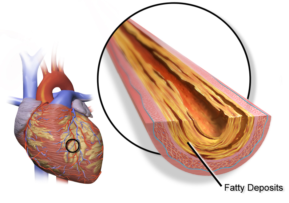
Coronary Artery Disease
Coronary Artery Disease is the narrowing of or a blockage in the coronary arteries. When this narrowing occurs, the heart is not able to receive enough oxygenated blood. The lack of oxygenated blood may cause an individual to experience chest pain (angina pectoris). This pain usually occurs when the heart’s demand for oxygen is greater than the supply, especially during times of physical or emotional stress. As an artery continues to narrow, blood supplied to the heart muscle decreases further, causing more chest pain. If the artery becomes completely blocked or too narrow to supply enough oxygenated blood to the heart muscle, it leads to what is known as a heart attack or myocardial infarction.
Heart Valves
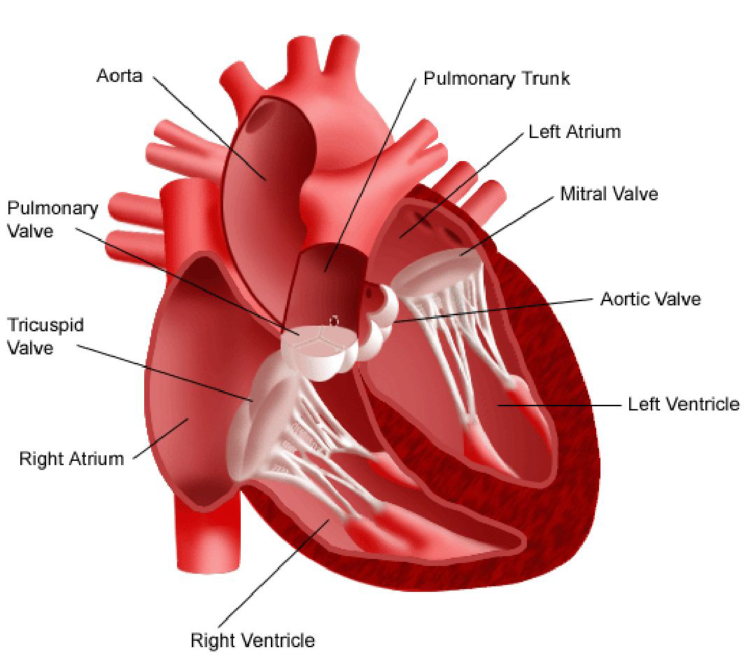
A heart valve is a ring-like structure with smooth leaflets or cusps. These cusps serve to control the flow and direction of blood as it passes through the heart chambers and out into the body. The heart has four main valves:
- Tricuspid Valve: Between the right atrium and the right ventricle.
- Mitral Valve: Between the left atrium and the left ventricle.
- Pulmonary Valve: Between the right ventricle and the lungs.
- Aortic Valve: Between the left ventricle and the body.
Valvular Heart Disease
The valves in the heart are normally thin, smooth leaflets that allow blood to flow through the heart chambers in a single direction. Heart valves may become diseased for a variety of reasons, including infection, rheumatic fever, birth defects, and old age. Heart valves may become stiff or calcified, causing a narrowing that prevents blood from flowing freely. This is called stenosis. A damaged valve may be thickened and scarred so that it does not open all the way. A stenotic valve hinders the flow of blood from one chamber to another because of the narrow opening. Another change that can occur in the valve is called insufficiency, which happens when the valve does not close all the way. This incomplete closure causes blood to flow backward from one chamber into another. You may hear your doctor call this backward flow of blood leaking or regurgitation.